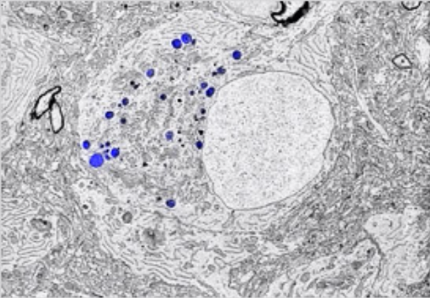
Cancer research is an important field in medicine which investigates the causes of cancer and develops methods for treatment. Given our limited knowledge of its development, progression and metastasis, cancer remains to be one of the most daunting challenges in medical research. Understanding these processes that happen on a nanoscale level within a single cell or a group of cells is essential to developing methods that can limit the spread of cancer to other parts of the body. Correlative light and electron microscopy (CLEM) a hybrid imaging technique that bridges this gap by combining the strengths of light microscopy and electron microscopy, has been gaining popularity in the field of cancer research.
What is CLEM?
Correlative Light and Electron Microscopy(CLEM) links the two imaging techniques; fluorescense microscopy (FM) and electron microscopy (EM). Fluorescense microscopes, also known as light microscopes, are typically used for observing the overall structure of cells. They allow for live cell imaging and the use of fluorescence markers to observe dynamic biological processes in real time. However, they are limited for observing the details of subcellular structures. On the other hand, electron microscopes have allowed researchers to get a better understanding of sub cellular structures, as they have a higher resolution and greater resolving power. The great potential of CLEM microscopy lies in the fact that it enables you to correlate the two different types of information on the exact same area of interest: cellular function from FM and ultrastructure from EM. This combination enables researchers to correlate molecular dynamics with high-resolution structural data. As a result, CLEM provides a more complete view of how cancer cells behave and change at both the structural and functional levels.
CLEM in Cancer Research
CLEM has become an important tool for cancer research with its abilities for researchers to study cancer cells in their natural state. It has been contributing in visualizing disease pathogenesis and mechanisms at the sub-cellular level, including how cancer cells invade surrounding tissues and migrate and spread to distant organs, the process known as metastasis. For instance, studies using CLEM have shown how cancer cells reorganize their cytoskeleton to invade surrounding tissues or evade the immune system.
One of the key contributions of CLEM in cancer research is its ability to offer new perspectives on cancer progression and metastasis, as CLEM allows scientists to directly observe changes in cellular structures that power tumor growth and the spread of cancer. In particular, CLEM has helped researchers to study how cancer cells break through the extracellular matrix (ECM), a network of proteins supporting cells and tissues in the body.
The Future of CLEM Technology
As imaging technology continues to advance, CLEM holds immense potential to revolutionize cancer research. Future advancements in both fluorescence microscopy and electron microscopy could lead to even more precise imaging, allowing scientists to trace cellular changes in more complex systems. Furthermore, CLEM’s ability to visualize cancer cells in their natural state could mean that it could become a key instrument in developing better cancer therapies that obstruct cancer progression and metastasis.
In short, novel technologies like CLEM will play a crucial role in scaling up and fastening cancer research, ultimately bringing us closer to finding a cure for cancer.
- Zinnia
Sources
- https://www.delmic.com/en/solutions/correlative-microscopy-cancer-research
- https://www.delmic.com/en/techniques/correlative-light-electron-microscopy
- https://www.shalom-education.com/courses/gcse-biology/lessons/cell-biology/topic/light-microscopes-and-electron-microscopes/
- www.capabilities.unsw.edu.au/correlative-light-and-electron-microscopy-clem-based-analysis-disease
- https://www.ncbi.nlm.nih.gov/pmc/articles/PMC8070945

